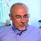
 Dr. László Mecseky
Surgeon/Traumatologist
Diabetic Leg Specialist
President of Diabetic Leg Alliance, Hungary
Founder of National Diabetic Leg Screening Program at Hungary
Results in the prevention and treatment of diabetes complications are still far from satisfactory. Statistics show a grim picture: one in every 10 people in Hungary may be affected on account of the spread of diabetes. However, we can safely say that nowadays with proper care and treatment every second amputation could be prevented.
What possibilities do we have to improve these grim figures?
- recognizing diabetes at its early stage and preventing complications
- ensuring complex treatment
- preventing the recurrence of complications
- improving patients’ cooperative skills
The situation is sometimes made even more complicated by misconceptions:
- acts of pessimism: “it will have to be amputated sooner or later anyway, it’s just a question of time”
- the other extreme is when the situation is belittled as if foot complications could be cured and prevented by taking medications only.
Numerous amputations are carried out because a lot of patients are unaware of the dangers their illness poses and do not dedicate sufficient attention and care to their condition. They fail to go to diabetic care regularly and it is only when foot wounds have already appeared, never before, that they see the local physician, usually with uncared-for wounds.
The illness means neuro-angiopathy, however, it is still very far from being an untreatable vessel structure.
Serious limb-threatening ischemia, in which invasive surgery or radiological methods are to be applied, account for a mere 10% of the cases.
Amputees are automatically diagnosed with “arteriosclerosis” in the medical records, a term virtually applicable for anything, and the real reasons remain unrevealed.
“Clear neuropathy” is characteristic for at least 70% of diabetic patients and in this group no amputation should ever be carried out. The timely diagnosis of neuropathy is always crucial as it is its consequences that lead to tragedies.
Motor neuropathy leads to muscular atrophy, as a result of which the foot deforms. Pathologic pressure points appear on the sole where ulcers can easily appear. (Figure 1)
Because of autonomic neuropathy, keratoses of incredible size can appear, which may lead to further deep tissue damages. (Figure 2)
Figure 1: Plantogram indicates points of overpressure where keratosis appeared.
Figure 2: Ulcer under keratosis
2013/4 37
Autonomic neuropathy is responsible for inadequate perspiration, as a consequence of which the skin becomes dry and vulnerable. The latter phenomenon is further complicated by the fact that diabetic patients’ skin does not contain hyaluronate, which plays an important role in the flexibility, conditioning and resistance of the skin.
The most dangerous neuropathy and the curse of diabetes is sensory neuropathy since irreversible processes resulting from a delay in treatment are due to insensitivity, i.e. the patient’s incapability to feel pain.
In light of what has been mentioned so far it is easy to understand why inadequate foot care and unsuitable shoes account for 70% of the amputation risk factor.
If the existence of neuropathy has been proved with diapason test, tests for heat sensation or the application of monofilament, providing the patient with neuropathy protecting shoes are of utmost urgency as well as ensuring proper skin protection and nail care.
One of the reasons of unnecessary amputations is that all these measures are taken too late and the prevention period is skipped entirely. I had a patient who had already been through all kinds of “juggling” with their sole ulcer, yet, still had not been provided with information about even the most basic pressure relieving methods. It is most unfortunate that even non-professional events dedicated to providing information to diabetic patients fail to introduce the proper tools, shoes and ortheses to the patients because they are supported by the National Health Care Fund. In light of this it is no wonder that patients are occasionally provided with completely unsuitable medical aids.
What is of primary importance in the crucial preventive period is the application of preventive shoes, tailor-made insoles, gels containing zinc-hyaluronate for skin care and the in-time commencement of anti-onychomycosis and anti-dermatomycosis treatments. By observing the abovementioned advice the illness could be prevented in its very root.
The seemingly banal callosities at the tip of the toes, the fissures on the sole and lesions in the thickened area around the nails lead to serious consequences, the end of which, if not treated adequately, is amputation. As an aggravating factor, all these are accompanied with the patient’s procrastinating behaviour due to their loss of sensation. (Figure 3)
If the septic process fails to get discovered in time and is not treated professionally (meaning the removal of the source, not amputation!), the process can spread between the deep tissues of the foot unnoticed for a long time and when the inflammation appears above the foot, saving the limb is far more difficult.
However, there are certain warning signs the evaluation of which is crucial even at the general practitioner’s. One such sign is sudden monolateral swelling of the foot or tarsus.
Whether there are deep tissue septic causes in the compartment syndrome or it is Charcot osteoarthropathia resulting in the collapse of the joints, the most important thing is to ensure that the patient should not stand on their feet at all until the nature of the process has been identified unequivocally lest they should aggravate the situation with every step. If the process is purulent, every step taken squeezes a portion of purulent matter into the ascending lymphatic system, in the case of Charcot foot the joint structures are crumbled as if they were being crushed in a mortar.
Figure 3: Callosities ate the tip of the toe, pathological nail
Figure 4: Longitudinal surgical exposure of compartment syndrome and healed status
38 2013/4
It is not mere coincidence that these two diabetes complications are called catastrophic medical conditions. Sadly enough, medical treatment is also often disastrously procrastinative since septic expansive deep tissue procedures require wide, longitudinal surgical exposure, in the case of which fear of extensive wounds should not hinder the surgeon’s work. It is like excising a tumour: either we remove it from the intact area or we have not made any progress towards healing. Therefore what we need is not minor incisions but a thorough, or even “brutal”, exposure all along the spreading of the purulent process. (Figure 4)
After saving the limb with this 2-minute long ambulatory intervention, we may think about continuing the treatment, using modern open wound treatment, the application of gels containing zinc-hyaluronate to facilitate the early healing of long scars or the use of pressure relieving and –distributing total contact insoles and proper stable protective shoes capable of holding it. (Figure 5)
After proper treatment patients can walk again in 2-3 weeks’ time. However, if the patient with compartment syndrome does not receive professional help in time or exposure is delayed by the doctor waiting for the proper conditions of anaesthesia, the limb is lost. In the deep tissues of the sole and in the areas surrounded by tight fascias the purulent processes may result in a vicious circle, which results in the necrosis of the toes of the affected area. Although the thus formulated picture raises the possibility of ischemia, unfortunately disastrous misdiagnoses are quite common. Yet, it is anything but ischemia! The lack of oxygen in the affected area is solely caused by local processes, i.e. the increasing local pressure which can immediately be cured once the pressure ceases. The diabetic foot is characteristically insensitive anyway and in case of an advanced compartment syndrome this insensitivity might even increase, therefore there is no need for anaesthesia. Yet, if there are still unexplored recesses remained, the treatment naturally has to be continued in a surgery with anaesthesia. I. v. antibiotics might be required, therefore hospitalization might be absolutely necessary for a few days.
If the cause of monolateral swelling of the foot or tarsus is not purulence, the problem is called Charcot foot. In this case urgency is required in establishing diagnosis as soon as possible, relieving the area of pressure and superannuation. (Figure 6)
It is a serious everyday problem that these patients carry out their visits to departments of rheumatology, orthopaedics, dermatology, diabetology, traumatology and the physiotherapist ON FOOT until the proper diagnosis is established.
With physical examination it is easily palpable that the tarsus bones are like ice cubes in a nylon sack. Certainly, the patient feels nothing of all that, therefore neither they nor their environment take the illness seriously.
What to do: immediate pressure relieving with two crutches, a walker, then RTG as well as referral of the patient to specialists of diabetes complications. Further diagnostic steps, such as CT or MR are special competences already.
Treatment is usually conservative in nature: compression bandage, plaster cast, orthesis, occasionally surgery because of osteomyelitis resulting from procrastination or in cases where the foot structure has been wrongly fixed. Compression bandaging and plaster casting carried out with insufficient practice are usually accompanied by skin damages of various degrees.
Figure 5: Protecting shoes, insoles
Figure 6: Charcot foot
2013/4 39
Their number can considerably be reduced with the in-time application of gels containing zinc-hyaluronate. The “fixing” of the collapsed foot structure, i.e. osteosynthesis is often accompanied by further complications, the already osteoporotic bones do not agree with metal implants. If we knew the illness already at the beginning – even an easy routine piezometry at the sole might indicate this when making protective shoes –, there would still be alternatives. The aim: stable foot synostosis in a good position, which can be used in rolling sole shoes with tailor made insoles. In other words, do not try to exercise the “exploded” joints as that will surely lead to instability and walking disability.
Fortunately, Charcot foot is rare, few people are exposed to it. However, septic complications appearing on the toes or acras are common, therefore the family doctor has great responsibility in the professional in-time treatment of these symptoms. If the source is found in time, whether at the nail edges, under the nail or at the bone of the distal phalanx, removing the source is easy, there is no need for amputating the toe. A minor ostitis shall never lead to e.g. the loss of a big toe!
The causes – e.g. inadequate carbohydrate metabolism – always have to be clarified!
Amputation should never be the first option!
Fortunately Diabetes Foot Outpatient Care Centres are spreading in Hungary, providing proper follow-up treatment to patients undergoing surgery, modern treatment of chronic wounds and preventive measures against recidivation. More and more of them provide modern atraumatic “medical chiropody” as a part of the complex treatment, in which the exploration of septic sources is also an important task. (Figure 7) Figure 7: Medical chiropody
The latter predominantly means dry machine-assisted foot care under minor theatre circumstances with medical supervision. However, the establishment of outpatient care centres providing complex treatment is still hindered by fiscal considerations, often even medical aids are not provided in time. Yet, development is continuous despite the grave economic circumstances, an increasing number of modern dressing materials are becoming state-supported. Most of these can be combined with gels containing zinc-hyaluronate to create a flexible, quick-healing scar and to prevent deep fissures causing prolonged wound healing. With the end of discharging the application of dressing materials can also be suspended since the application of gels containing zinc-hyaluronate twice a day creates a proper protective layer on the wound.
It is important to know that without modern open wound treatment the diabetes foot cannot be healed and long-healing ulcers deprive patients of their chance to live a decent life.
Numerous professional forums indicate an unequivocal consensus that billions of forints can be saved with professional treatment and the saving of limbs saves lives as well. If we can reduce the number of recidivas, perspectives of recovery become much sounder. The supply of modern medical aids and patient information, the enhanced understanding of both the patient and their environment all contribute to the decrease in the number of recurrences. By wearing permanent medical aids (protective shoes with tailor made insoles), replacement of missing constituents of the skin (zinc-hyaluronate, urea), the application of compression treatment (rubber bandage), ensuring a well-balanced carbohydrate metabolism and applying medicine for neuropathy, the risk of the recurrence of complications is minor.
However, in order to achieve this it is not enough to see a specialist of diabetes complications (Diabetes Foot Outpatient Care Centre) when the problem is already severe because at that stage the only thing the doctor can do is mitigate the already existing damage.
Regular monitoring of diabetic patients’ feet, upgrading of medical aids and continuous skin and nail care are necessary to save the limbs!
Let’s not forget that after amputations the risk of losing the opposite limb is high (60% in 5 years’ time) and 40-70% of the amputees die within 5 years.
Literature: in the editorial office
40 2013/4